Cross Section Of A Compact Bone / Bone Marrow Transplant: Procedure & Cost | Narayana Health : This model shows a cross section of compact bone.
Cross Section Of A Compact Bone / Bone Marrow Transplant: Procedure & Cost | Narayana Health : This model shows a cross section of compact bone.. □ on examining a cross section of any bone, it is composed of two kinds of bony tissue: Although compact bone is made up of haversian systems, it is almost solid. The basic unit of structure in this type of bone is the haversian system, or osteon. Magnification view of compact bone tissue. Remodeling allows the body to fix damaged sections, reshape the skeleton during growth, and regulate calcium levels.
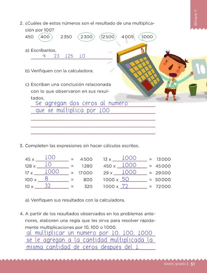
The spongy and compact bone tissue in the cross section of a skull bone. Compact bone, also known as cortical bone, is a denser material used to create much of the hard structure of the skeleton. The first, outermost layer of membrane (besides periosteum) serves to. Concentric layers of bone cells (osteocytes) and bone matrix surround. Remodeling allows the body to fix damaged sections, reshape the skeleton during growth, and regulate calcium levels.
Bone Anatomy | Ask A Biologist from askabiologist.asu.edu Structures and bone areas in column b, and use them to color the coding. Bone tissue epiphysis (end) of a long bone longitu. Compact bone, makes up the dense material in a long section of a bone. Sclerostin inhibits bone formation mostly by antagonizing lrp5/6, thus inhibiting wnt signaling. The basic unit of structure in this type of bone is the haversian system, or osteon. The osteon consists of a central canal called the osteonic (haversian) canal, which is surrounded by concentric rings (lamellae) of matrix. In the center of each osteon is the central canal, a space that houses blood vessels and nerves that supply bone. As seen in the image below each osteon is also composed of a number of different cells responsible for the maintenance of the bones, including osteocytes and osteoblasts.
A diagrammatic view of a cross section of bone.
□ on examining a cross section of any bone, it is composed of two kinds of bony tissue: Concentric layers of bone cells (osteocytes) and bone matrix surround. Magnification view of compact bone tissue. □ compact tissue, it is dense in texture and it is always the osteoblasts form a collar of compact bone around the diaphysis. Compact bones make up 80 percent of the human skeleton; Compact bone is very different from the other tissues you have seen. A diagrammatic view of a cross section of bone. In the center of each osteon is the central canal, a space that houses blood vessels and nerves that supply bone. (b) in this micrograph of the osteon, you can clearly see the concentric lamellae and central canals. The first, outermost layer of membrane (besides periosteum) serves to. The spongy and compact bone tissue in the cross section of a skull bone. Compact bone consists of closely packed osteons or haversian systems. This is a short tutorial using blender 2.8 that shows how to create a bone cross section and using images to create the textures.
Observe that the matrix of the bone is deposited in concentric layers that are called lamellae (5). The remainder is spongelike cancellous bone. Hope you enjoy and please. Cross section of the compact bone. Structures and bone areas in column b, and use them to color the coding.
Bone Marrow Stimulating Medications Boost Blood Counts from fthmb.tqn.com Between the rings of matrix, the bone cells (osteocytes) are located in spaces called lacunae. This is a short tutorial using blender 2.8 that shows how to create a bone cross section and using images to create the textures. In the last decade, considerable technological improvements have been made to repair damaged bones and tissue, such as bone cross sections with implants for microscopic examinations. Gross observation of bone in cross section shows dense areas generally without cavities—corresponding to compact bone—and close gross examination of a thick section of dried bone illustrating the cortical compact bone and the lattice of trabeculae in cancellous bone at the. Compact bone (cross section of dried bone). Select different colors for the. Compact bone consists of closely packed osteons or haversian systems. The remaining material is mostly collagen.
This is a short tutorial using blender 2.8 that shows how to create a bone cross section and using images to create the textures.
(micrograph provided by the regents of university of michigan. Although compact bone is made up of haversian systems, it is almost solid. These are abundant and characteristic of compact bone. Remodeling allows the body to fix damaged sections, reshape the skeleton during growth, and regulate calcium levels. Cross section of ground compact bone. Bone must be decalcified (by exposure to strong acids) so it can be cut into thin sections. Your microscopic section compact bone stock images are ready. In the last decade, considerable technological improvements have been made to repair damaged bones and tissue, such as bone cross sections with implants for microscopic examinations. 850 x 560 png 177kb. (b) in this micrograph of the osteon, you can clearly see the concentric lamellae and central canals. Magnification view of compact bone tissue. The remaining material is mostly collagen. □ on examining a cross section of any bone, it is composed of two kinds of bony tissue:
In the center of each osteon is the central canal, a space that houses blood vessels and nerves that supply bone. Your microscopic section compact bone stock images are ready. Compact bone, also known as cortical bone, is a denser material used to create much of the hard structure of the skeleton. The innermost layer of membrane is made up of. Observe that the matrix of the bone is deposited in concentric layers that are called lamellae (5).
Labeled Histology Slides | Histology slides, Anatomy and ... from i.pinimg.com Compact bone consists of closely packed osteons or haversian systems. There are trabeculae in spongy bone which gives its sponge like appearance. Your microscopic section compact bone stock images are ready. This makes it very dense, so it has a high mass. These are mostly compacted bone with little marrow and include most of the bones in the limbs. A this cross sectional view of compact bone shows the basic structural unit the osteon. Structures and bone areas in column b, and use them to color the coding. This is a short tutorial using blender 2.8 that shows how to create a bone cross section and using images to create the textures.
Compact bone (cross section of dried bone).
The osteon consists of a central canal called the osteonic (haversian) canal, which is surrounded by concentric rings (lamellae) of matrix. They build the entire picture, improve your understanding, consolidate the information and facilitate recall. Observe that the matrix of the bone is deposited in concentric layers that are called lamellae (5). The innermost layer of membrane is made up of. □ compact tissue, it is dense in texture and it is always the osteoblasts form a collar of compact bone around the diaphysis. At the same time, the cartilage in the center of the diaphysis begins to disintegrate. A cross section of a compact bone shows concentric circles called lamellae. Most bones contain both compact and spongy bone. This model shows a cross section of compact bone. There are trabeculae in spongy bone which gives its sponge like appearance. Select different colors for the. Compact bone is very different from the other tissues you have seen. Compact bone (cross section of dried bone).
In the center of each osteon is the central canal, a space that houses blood vessels and nerves that supply bone cross section of a bone. □ compact tissue, it is dense in texture and it is always the osteoblasts form a collar of compact bone around the diaphysis.

/compact-spongy-cancellous-bone-cross-section-of-a-long-bone-shows-compact-bone-spongy-cancellous-bone-and-marrow-cavity-139820224-57ed42be5f9b586c35d79464.jpg)